
Yes, minerals have a say in preservation of environmental DNA
- Nov 22, 2023
- 2 min read
Super constructive collective efforts paved the way for our new study published in "Environmental DNA." We introduce mineralogy and interfacial geochemistry to the field of ancient environmental sedimentary and show how mineral topography and charge density can control DNA preservation.
Take home:
Sediment mineralogy matters for DNA taphonomy and preservation. Knowledge of the mineralogical composition of a sediment and the environmental conditions can help assess if a sediment can have stored extracellular DNA and to what extent the DNA would be preserved.


The AFM image is 2 um across. The white bands are plasmid/circular DNA. Toosin at the AFM mounting a piece of calcite for his force measurements. Schematic of the AFM tip and the laser feedback system.
The long process short:
Léa was very instrumental in facilitating the paper. She paved the way for the AFM work and I still remember when she first showed me the AFM results: Circular DNA conformed to the angular step edges of the calcium carbonate surface. Colin did MD simulation and the results were nicely overlapping with our experimental data.
One thing troubled us: -Why did the DNA fragment after binding to the calcite surface?
Colin ran some more simulations and I ran some more liquid cell AFM experiments and we finally realized that that the binding to the mineral surface induce a loss of H bonds in the helix causing a breakdown of the helix. Marina made great efforts in extracting DNA adsorbed to calcite and could confirm fragmentation. Toosin consolidated Léa´s AFM work on the dependence of salinity and ionic strength for DNA - mineral interaction as he used chemical force spectroscopy to show how solution composition can influence the DNA-mineral interaction strength. As part of Jaime's MSc thesis he helped reproducing the AFM image data. Matthew, great discussions as always and thanks for bringing Colin over!
Thank you VILLUM FONDEN for seeing the merits in our type of work. Having a video rate atomic force microscope (Cypher VRS) in the group help us break some ground.
You can read the full study here:
It is open access so crack on.

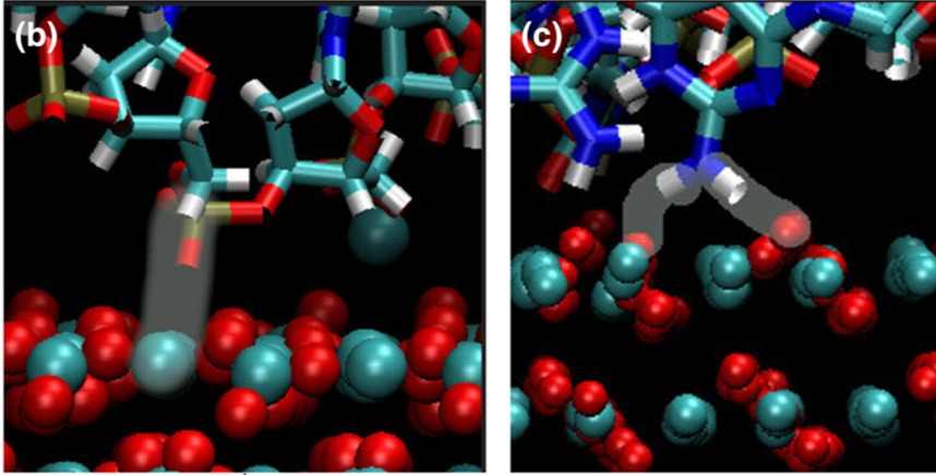

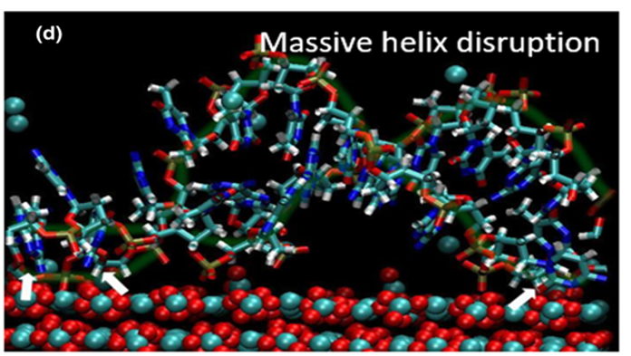
Check out Colins work using molecular dynamics simulations (Fig. 3 and 4). He shows that DNA binding at the step edges can cause massive helix disruption.


Comments